
Dr. Alessandro Giardini
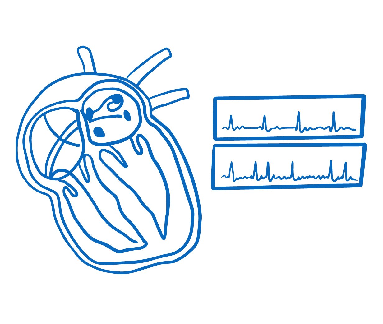
An electrophysiology (EP) study is a specialised procedure used to investigate abnormal heart rhythms (arrhythmias) in children. If the source of the rhythm problem is identified during the study, it can often be treated at the same time using a technique called catheter ablation. Together, these procedures offer the possibility of a lasting cure for many types of arrhythmia, potentially removing the need for long-term medication.
The procedure is carried out in a dedicated electrophysiology laboratory within the hospital. Your child will receive a general anaesthetic so that they remain completely asleep and comfortable throughout. Once the anaesthetic has taken effect, thin, flexible tubes called catheters are inserted through blood vessels, usually in the groin, and carefully guided to the heart under X-ray imaging. Small electrodes at the tips of these catheters record the heart's electrical signals from inside, allowing Dr Giardini's team to build a detailed map of the electrical pathways and pinpoint exactly where the rhythm disturbance originates.
During the study, the team may use gentle electrical impulses delivered through the catheters to reproduce your child's abnormal rhythm in a safe and controlled environment. This step is important because it confirms the precise mechanism of the arrhythmia and identifies the area of heart tissue responsible. Common conditions investigated in this way include supraventricular tachycardia (SVT), Wolff-Parkinson-White syndrome, and arrhythmias that can develop after surgical repair of congenital heart defects.
If the source of the arrhythmia is located and ablation is appropriate, a specially designed catheter is positioned over the abnormal area. A small amount of energy is then applied to that precise spot to permanently alter the tiny piece of tissue causing the problem. Two types of energy are used: radiofrequency (RF) ablation, which uses controlled heat, and cryoablation, which uses targeted cold. Both are highly effective, and the choice depends on the location of the abnormal tissue and what is safest for your child. Modern three-dimensional mapping systems allow the team to target the area with great accuracy while keeping radiation exposure to a minimum.
The procedure typically takes two to three hours, though this can vary depending on the complexity of the arrhythmia. Throughout, a paediatric cardiac anaesthetist monitors your child continuously, and the team will keep you updated on progress. Once the procedure is complete, the catheters are removed and firm pressure is applied to the insertion site. Your child will be taken to a recovery area where you will be able to be with them as they wake up. A pressure bandage is placed over the groin, and your child will need to lie still for a few hours afterwards. Most children are able to go home on the same day or the following morning and can return to normal activities within about a week.
Dr Giardini will discuss the results with you as soon as the procedure is finished. If the ablation is successful, most children will no longer need medication to control their arrhythmia. If you would like to discuss whether an EP study or ablation may be appropriate for your child, please do not hesitate to contact Dr Giardini's team to book an appointment.