Marfan Syndrome in Children and Adolescents
Written by Dr Alessandro Giardini, Consultant Paediatric and Adolescent Cardiologist in London, UK
What Is Marfan Syndrome?
Marfan syndrome is a genetic condition that affects connective tissue, the material that holds the body together and gives strength and flexibility to blood vessels, bones, ligaments, muscles, and skin. Because connective tissue runs throughout the body, Marfan syndrome can affect many different systems, including the heart and blood vessels, the skeleton, the eyes, the lungs, and the skin.
The condition takes its name from Antoine Marfan, a French doctor who first described it in 1896. It ranks among the most common inherited connective tissue disorders, affecting roughly 1 in 3,000 to 5,000 people worldwide. It occurs equally in boys and girls and across all ethnic backgrounds.
The most important concern with Marfan syndrome is the effect it has on the aorta, the main blood vessel that carries blood away from the heart. The walls of the aorta can weaken, causing it to widen (a condition called an aneurysm) or, in serious cases, to tear (known as an aortic dissection). Before modern treatments existed, these heart and blood vessel problems significantly shortened life expectancy. Today, with early diagnosis, regular monitoring, medication, and surgery when needed, people with Marfan syndrome can expect to live a near-normal lifespan.
Many features of Marfan syndrome become more noticeable during the teenage growth spurt, which means doctors can find the condition hard to diagnose in younger children. If a family history exists, doctors can monitor children from birth. If there is no family history, the diagnosis may not come until a child is older. However once the doctors recognises it, effective management can begin straight away. As a paediatric cardiologist I have cared for many children and families living with Marfan syndrome and understand how important it is to have clear, trustworthy information when you face this diagnosis.
What Causes Marfan Syndrome?
A change (mutation) in a gene called FBN1 on chromosome 15 causes Marfan syndrome. This gene provides the instructions for making a protein called fibrillin-1. Fibrillin-1 is one of the main building blocks of the tiny fibres (microfibrils) that give connective tissue its strength and elasticity. When the FBN1 gene changes, the fibrillin-1 protein stops working properly, and the connective tissue throughout the body becomes weaker and stretchier than it should.
The mutation also drives increased activity of a signalling system called the TGF-beta pathway, which plays a role in how tissues grow and repair themselves. This overactivity contributes to many of the features of Marfan syndrome, particularly the widening of the aorta.
Marfan syndrome follows an autosomal dominant pattern of inheritance, meaning that a single copy of the altered gene is enough to cause the condition. If a parent has Marfan syndrome, each of their children has a 1 in 2 (50 per cent) chance of inheriting it. About 75 per cent of people with Marfan syndrome have inherited the gene change from an affected parent even though in some cases nobody has yet diagnosed the parent. In the remaining 25 per cent, the gene change occurs for the first time in the child (a de novo mutation), with no family history of the condition.
As with many genetic conditions, Marfan syndrome varies considerably from person to person. Two people in the same family carrying the same gene change may experience the condition quite differently: one mildly, the other more significantly. This variability can make diagnosis challenging, but it also means that having Marfan syndrome does not automatically predict how severe a child’s symptoms will become.
What Are the Symptoms of Marfan Syndrome?
Marfan syndrome can affect several parts of the body. Many features start mild and become more apparent during adolescence, though some may show from birth. Not every child will have all of these features.
Heart and blood vessels
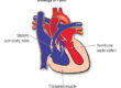
This is the most important area. The aorta and particularly the section closest to the heart called the aortic root tends to widen gradually over time. Without monitoring and management, this widening can lead to an aortic aneurysm (a dangerous bulge in the wall) or an aortic dissection (a tear in the wall), both of which constitute medical emergencies. The mitral valve, which sits between the two left chambers of the heart, also commonly develops problems. Mitral valve prolapse, a condition where the valve flaps become floppy and do not close properly occurs in many children with Marfan syndrome and can sometimes cause the valve to leak. Less commonly, the aortic valve or the tricuspid valve may also cause concern.


Skeleton
Children with Marfan syndrome often grow taller than expected for their age, with long arms, legs, fingers, and toes. Doctors describe the long fingers as arachnodactyly (literally “spider fingers”). Other skeletal features can include a sunken chest (pectus excavatum) or a protruding chest (pectus carinatum), curvature of the spine (scoliosis), very flexible joints (joint hypermobility), flat feet, and a high-arched palate (roof of the mouth) that may crowd the teeth. These features tend to become more noticeable during the growth spurt of puberty.
Eyes
Eye problems form a hallmark of Marfan syndrome and one of the features that help doctors make the diagnosis. The most characteristic finding is dislocation of the lens of the eye (known as ectopia lentis), which occurs in about 60 per cent of people with Marfan syndrome. The lens shifts out of its normal position because the weakened tiny fibres can no longer hold it in place. Short-sightedness (myopia) is very common, often more severe than usual. Children with Marfan syndrome also face an increased risk of retinal detachment, glaucoma, and early cataracts.
Lungs
The lungs can develop problems in some children. The risk of spontaneous pneumothorax rises. A pneumothorax occurs when air leaks out of the lung and into the space between the lung and the chest wall, causing the lung to partially collapse. It can cause sudden chest pain and breathlessness and needs prompt medical attention.
Skin
Stretch marks (striae) may appear even without significant weight gain, often on the lower back, hips, or shoulders. The skin may bruise more easily than normal.
Nervous system
In some cases, the covering of the spinal cord (the dura) can stretch and weaken, a condition called dural ectasia. This most commonly affects the lower spine and may cause lower back pain or leg pain, though many people with dural ectasia notice no symptoms at all.
How Is Marfan Syndrome Diagnosed?
Diagnosing Marfan syndrome involves bringing together information from a physical examination, heart scans, eye tests, family history, and genetic testing. Doctors use a set of internationally agreed criteria known as the revised Ghent nosology to reach the diagnosis.
Physical examination
A specialist, usually a paediatric cardiologist and/or clinical geneticist, carries out a detailed examination, looking at the child’s height, body proportions, chest shape, spine, joints, and facial features. A scoring system adds up how many of the typical skeletal, skin, and other features the child shows. Because Marfan syndrome can prove difficult to diagnose in younger children whose features are still evolving, a cardiologist experienced with inherited connective tissue disorders brings particular value.
Echocardiogram (heart ultrasound)
This painless scan uses sound waves to create a picture of the heart. It measures the size of the aortic root, checks the heart valves (particularly the mitral and aortic valves), and assesses how well the heart pumps. This stands as the single most important test in monitoring Marfan syndrome, and doctors typically perform it at diagnosis and then at least once a year.
Eye examination
A thorough eye examination by an ophthalmologist is essential. The specialist checks for lens dislocation (which may need the pupils to dilate for a clear view), short-sightedness, and any signs of retinal problems or glaucoma.
Genetic testing
A blood test can identify changes in the FBN1 gene. Genetic testing finds the mutation in about 95 per cent of children with Marfan syndrome. This helps not only to confirm the diagnosis but also to test other family members and plan for the future. However, doctors can also make a diagnosis of Marfan syndrome on clinical grounds alone if enough characteristic features are present.
Additional imaging
CT or MRI scans may look at the full length of the aorta and other blood vessels, particularly in older children and adolescents. Doctors generally prefer MRI in children because it avoids radiation. They also use these scans to check for dural ectasia if the child has symptoms of back or leg pain. X-rays of the spine and pelvis may assess scoliosis.
Family assessment
Because 75 per cent of Marfan syndrome cases run in families, first-degree relatives (parents, brothers, and sisters) of a diagnosed child should receive an assessment. This can involve a clinical examination, an echocardiogram, and genetic testing if the family’s specific gene change is known.
What Treatments Are Available?
No cure for Marfan syndrome exists, but it ranks among the most treatable genetic conditions. With the right management, the vast majority of children with Marfan syndrome go on to live long, active, and fulfilling lives.
Medication
The main aim of medication is to reduce the strain on the aorta and slow down any widening. Beta-blockers (such as atenolol) are the most commonly used drugs. They work by slowing the heart rate and lowering blood pressure, which reduces the force with which blood pushes against the aortic wall with each heartbeat. Doctors also widely use angiotensin receptor blockers (ARBs), particularly losartan. Research has shown that losartan directly affects the TGF-beta signalling pathway that drives Marfan syndrome, and it may offer additional protection to the aorta beyond blood pressure lowering alone. Some children take a combination of both a beta-blocker and an ARB, particularly if the aorta is enlarging more quickly than expected. As your child grows, the team will need to adjust the doses.
Regular monitoring
Lifelong surveillance forms a cornerstone of Marfan syndrome management. Most children have an echocardiogram at least once a year to track the size of the aortic root and check the heart valves. If the aorta is getting bigger or there are other concerns, the team may arrange scans more frequently. Eye examinations should also take place regularly, typically at six months of age, at three years, before school entry, and then every one to two years throughout childhood. The team should check the spine at each visit, particularly during the rapid growth of puberty, as scoliosis can progress quickly during this time. I work with each family to develop a monitoring plan that is thorough but also practical, keeping the number of hospital visits as manageable as possible.
Surgery
Doctors recommend preventive surgery on the aorta when the aortic root reaches a size at which the risk of a tear becomes significant. For most people with Marfan syndrome, this threshold sits at an aortic root diameter of 5.0 cm, though the team may consider surgery earlier (at 4.5 cm) if additional risk factors exist, such as a family history of aortic dissection, rapid growth of the aorta, or a planned pregnancy. The most commonly performed operation is a valve-sparing aortic root replacement, where the surgeon removes the weakened section of the aorta and replaces it with a synthetic tube while keeping the patient’s own aortic valve. This avoids the need for lifelong blood-thinning medication that a mechanical valve replacement would require. Planned surgery carries a very low risk at experienced centres.
The child may also need surgery for other reasons, including repair or replacement of a leaking mitral valve, treatment of a dislocated lens, repair of a detached retina, or correction of severe scoliosis.
Eyes
Glasses or contact lenses can correct short-sightedness. If the lens dislocates significantly, the surgeon may need to remove it and replace it with an artificial lens. Any sudden changes in vision with flashes of light, new floaters, or a shadow across the vision should prompt urgent assessment, as they may indicate retinal detachment.
Bones and joints
A child with scoliosis may need bracing or, in more severe cases, surgery. Supportive insoles can often manage flat feet. Dental crowding and a high-arched palate may require orthodontic treatment. Physiotherapy can help with joint pain and hypermobility.
Exercise and activity
Staying active matters for children with Marfan syndrome, both for physical health and emotional wellbeing. Low to moderate-intensity exercise works well. Activities such as swimming, cycling, walking, golf, doubles tennis, and gentle hiking are generally safe. The key principle is that your child should feel comfortable enough to hold a conversation while doing the activity. As a specialist with a particular interest in exercise physiology in children with heart conditions, I help families navigate these decisions so that children can enjoy staying active while remaining within safe limits.
Families should steer children away from competitive and contact sports (such as football, rugby, and basketball), heavy weight-lifting, and high-intensity endurance activities (such as long-distance running or competitive rowing). These put sudden or sustained high pressure on the aorta. For children aged 10 and under with mild aortic findings, restrictions are generally less strict, and most recreational sports can work well. For children over 10, guidance depends on the size of the aorta and families should discuss it with the cardiologist.
Emotional and psychological support
Living with a lifelong condition that affects appearance and limits some activities can feel challenging, particularly during the teenage years. Children with Marfan syndrome may grow noticeably taller and thinner than their peers, which can attract unwanted attention. School staff should know about the condition. Psychological support and counselling can help greatly, and connecting with other young people who have Marfan syndrome, through organisations like the Marfan Foundation or the Marfan Trust, can make a real difference.
Team approach
Because Marfan syndrome affects so many body systems, care works best when a team of specialists coordinates it. This typically includes a paediatric cardiologist, cardiac surgeon, clinical geneticist, ophthalmologist, orthopaedic surgeon, and dentist or orthodontist. A lead clinician who keeps the big picture in view helps ensure that all aspects of care join up properly.
Frequently Asked Questions
What is Marfan syndrome and how common is it?
Marfan syndrome is a genetic condition that weakens the connective tissue throughout the body, affecting the heart and blood vessels, skeleton, and eyes in particular. It affects roughly 1 in 3,000 to 5,000 people and occurs equally in boys and girls. A change in a gene called FBN1 causes the condition, and most people inherit it from a parent, though in about 1 in 4 cases it occurs for the first time with no family history.
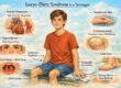
How is Marfan syndrome different from Loeys-Dietz syndrome?
The two conditions share some features, especially widening of the aorta. However, important differences exist. Lens dislocation is a hallmark of Marfan syndrome but does not occur in Loeys-Dietz syndrome (LDS). Conversely, LDS features widely spaced eyes, a split uvula or cleft palate, and twisted arteries — features that Marfan syndrome does not typically produce. In LDS, the aorta can tear at smaller sizes, so doctors recommend surgery at a lower threshold. Different genes cause the two conditions, and each requires a slightly different approach to monitoring and treatment.
What is the life expectancy for a child diagnosed with Marfan syndrome today?
This is understandably one of the first questions families ask, and the answer is very reassuring. Before modern treatments existed, life expectancy fell significantly short of normal. Today, with early diagnosis, regular monitoring, medication, and timely surgery when needed, life expectancy for people with Marfan syndrome has much improved and can sit close to that of the general population. The Marfan Foundation regularly hears from community members in their 60s, 70s, and even 80s.
Can my child still play sport and be active?
Yes! Staying active matters and doctors encourage it for your child’s overall health. Low to moderate-intensity activities such as swimming, cycling, walking, golf, gentle hiking, and doubles tennis are generally safe. The guiding principle is that your child should feel able to talk comfortably during the activity. Families should avoid competitive and contact sports, heavy weight-lifting, and high-intensity endurance exercise. For younger children (under 10) with only mild aortic findings, restrictions are generally less strict. Your child’s cardiologist will give personalised guidance based on the size of their aorta.
How often will my child need check-ups and scans?
At a minimum, most children with Marfan syndrome have an echocardiogram (heart ultrasound) once a year. If the aorta is enlarging or there are other concerns, the team may arrange scans every six months or more frequently. Eye examinations should take place regularly throughout childhood. The team should check the skeleton (particularly the spine) at each visit, especially during the rapid growth of puberty. Your child’s specialist team will tailor the monitoring schedule to their individual needs.
When is heart surgery needed?
Not every child with Marfan syndrome will need surgery, but many will at some point in their lives. Doctors recommend preventive surgery when the aortic root reaches 5.0 cm, or at 4.5 cm if additional risk factors exist, such as a family history of aortic dissection or rapid growth of the aorta. Surgeons most commonly perform a valve-sparing aortic root replacement, which carries an excellent safety record at experienced centres. The team always discusses the timing carefully with the family.
Should my other children and family members be tested?
Yes. Because 75 per cent of Marfan syndrome cases run in families, parents and siblings of a diagnosed child should strongly consider assessment. This may involve a clinical examination, an echocardiogram, and genetic testing if the family’s specific gene change is known. Sometimes a parent discovers they have the condition only after their child receives a diagnosis. Finding affected family members early allows monitoring to begin before problems develop, which is one of the most valuable steps a family can take.
What should I do in an emergency?
Sudden severe pain in the chest, back, or abdomen, especially if it feels like a tearing or ripping sensation, may signal an aortic dissection, which is a life-threatening emergency. Call 999 immediately. Similarly, a sudden onset of blurred vision, flashing lights, or a shadow across the vision could indicate a retinal detachment, which needs urgent assessment by an eye specialist. Your child should carry a medical alert card or bracelet stating that they have Marfan syndrome, so that emergency teams know the diagnosis and can act accordingly.
Where can I find support and reliable information?
The Marfan Foundation (marfan.org) is the leading global organisation, offering family support groups, webinars, educational resources, and a help centre staffed by specialists. In the UK, the Marfan Trust (marfantrust.org) provides practical guidance, support networks, and a downloadable paediatric guide for families. The Marfan Association UK (marfan-association.org.uk) is another useful resource. Connecting with other families who understand the day-to-day realities of living with Marfan syndrome can help enormously, particularly for children and teenagers who may feel isolated or different from their peers. If you would like to discuss your child’s diagnosis or arrange an assessment, I will be happy to help.
Dr. Alessandro Giardini, MD, PhD
Written 30/03/2026